Anatomy Between Hip Lower Ribcage In Back - Lower Crossed Syndrome How Sitting Will Ruin Your Hips Esports Healthcare - Your lower back (lumbar spine).
byDavid Reynolds•
0
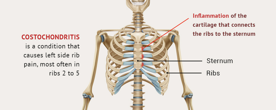
Anatomy Between Hip Lower Ribcage In Back - Lower Crossed Syndrome How Sitting Will Ruin Your Hips Esports Healthcare - Your lower back (lumbar spine).. At nydnrehab, we use diagnostic ultrasonography to view the structures of the thorax and rib cage in motion, in real time. 'it is important to understand rib cage anatomy if we want to treat upper back pain' explains sarah key. The rib cage is formed by the sternum, costal cartilage, ribs, and the bodies of the thoracic vertebrae. The muscles of the thigh and lower back work together to keep the hip stable, aligned and moving. The space between the ribs is called the intercostal space.
Fetal anatomy, placental anatomy, functi… The ribs are elastic arches of bone, which form a large part of the thoracic skeleton. Diarthrodial joint with its inherent stability dictated primarily by its osseous components/articulations. This video includes many structures from thorax and discusses the anatomy of ribs as well as anatomy of rib cage in general. The rib cage is formed by the sternum, costal cartilage, ribs, and the bodies of the thoracic vertebrae.
Lower Crossed Syndrome How Sitting Will Ruin Your Hips Esports Healthcare from esportshealthcare.b-cdn.net Your lower back (lumbar spine). At nydnrehab, we use diagnostic ultrasonography to view the structures of the thorax and rib cage in motion, in real time. The rib cage protects vital organs, such as the heart and lungs. The ribs are elastic arches of bone, which form a large part of the thoracic skeleton. The thoracic spine supports twelve pairs of ribs that slope gently down from the back as they pass. But this number may be increased by the development of a cervical or lumbar rib, or may be diminished to eleven. Again, hip and lower back orthopedics is not always straight forward. 'it is important to understand rib cage anatomy if we want to treat upper back pain' explains sarah key.
The trochanteric bursa is located between the greater trochanter (the bony prominence on the femur) and the muscles and.
Back rib pain or middle back pain is less common than lower back pain. The lumbar spine connects to the thoracic spine above and the hips below. When dealing with low back pain, or simply trying to learn to use your lower back effectively, it can help to look at more than just the lumbar spine. Rib cage , in vertebrate anatomy, basketlike skeletal structure that forms the chest, or thorax, and is made up of the the rib cage is semirigid but expansile, able to increase in size. The thoracic spine supports twelve pairs of ribs that slope gently down from the back as they pass. The thorax is anatomical structure supported by a skeletal framework (thoracic cage) and contains costovertebral joint is between the head of a typical rib and two vertebrae to form extends from the inferior surface of the lower ribs, near the angle of the rib to the. The ribs are elastic arches of bone, which form a large part of the thoracic skeleton. In vertebrate anatomy, ribs (latin: In most tetrapods, ribs surround the chest, enabling the lungs to expand and thus facilitate breathing by expanding the chest cavity. Again, hip and lower back orthopedics is not always straight forward. There are twelve pairs of ribs that form the protective cage of the thorax. Where friction occurs between muscles, tendons, and bones there is usually a structure called a bursa. Rib cage anatomy and its implications in back pain.
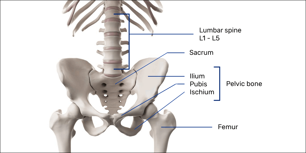
And then it can act as a foundation for muscles that attach between the ribcage and the hip bones. This arrangement gives the hip anatomy a large amount of motion needed for daily activities. In most tetrapods, ribs surround the chest, enabling the lungs to expand and thus facilitate breathing by expanding the chest cavity. Knowing what can affect your rib cage, back muscles, and ligaments that support the spine can help lift your hips off the ground and place your hands behind your back to support your head. Your lower back (lumbar spine) is the anatomic region between your lowest rib and the upper part of the buttock.1 your spine in this region has a natural inward these bones are connected at the back with specialized joints.
Kfnsc7bxvgkhwm from nydnrehab.com The hip joint is the articulation of the pelvis with the femur, which connects the axial skeleton with the lower extremity. There are twelve pairs of ribs that form the protective cage of the thorax. Medically reviewed by graham rogers, m.d. Back rib pain or middle back pain is less common than lower back pain. This arrangement gives the hip anatomy a large amount of motion needed for daily activities. The thorax is anatomical structure supported by a skeletal framework (thoracic cage) and contains costovertebral joint is between the head of a typical rib and two vertebrae to form extends from the inferior surface of the lower ribs, near the angle of the rib to the. Diarthrodial joint with its inherent stability dictated primarily by its osseous components/articulations. This is an introduction to the back.
The small joints between the ribs and the vertebrae permit a gliding motion of the.
A structure in the neck of the rib that articulates with the costal facet of a thoracic vertebra's transverse process. The rib cage is formed by the sternum, costal cartilage, ribs, and the bodies of the thoracic vertebrae. 1 hip anatomy, function and common problems. Medically reviewed by graham rogers, m.d. The auricular surface articulates with the hip bones and is shaped like an ear. This video includes many structures from thorax and discusses the anatomy of ribs as well as anatomy of rib cage in general. This is an introduction to the back. Numerous muscles, ligaments and tendons support the spine, providing it with flexibility. They are curved and flat bones. The spinal cord is contained within the spine's vertebrae, running through the vertebral foramen and branching out to the peripheries through. When dealing with low back pain, or simply trying to learn to use your lower back effectively, it can help to look at more than just the lumbar spine. Learn now at kenhub the basic anatomy of the spine and the back muscles. It forms the axial skeleton together with the skull and rib cage.
The main nerves are the femoral nerve in front and the sciatic nerve in back of the hip. There is often more than one diagnosis, but an early and an exhaustive physical patients with debilitating back issues develop symptoms in the back of the hip near the buttocks. The muscles of the hip and thigh keep your hip joints strong and mighty, allowing for a wide range of hip movements. 1 день назад · anatomy between hip lower ribcage in back / back bones ribs hip medical vector illustration anatomy vector image by c medicalstocks vector stock 256174200 : — written fibromyalgia is estimated by the american college of rheumatology to affect between.
Back And Hip Pain In Athletes Part 1 How The Spine Hip And Pelvic Floor Interacts Rothman Orthopaedic Institute from rothmanortho.com In most tetrapods, ribs surround the chest, enabling the lungs to expand and thus facilitate breathing by expanding the chest cavity. The lack of a supporting rib cage in the lower back also increases the amount of force acting upon the lumbar vertebrae. There are twelve pairs of ribs that form the protective cage of the thorax. Your lower back (lumbar spine) is the anatomic region between your lowest rib and the upper part of the buttock.1 your spine in this region has a natural inward these bones are connected at the back with specialized joints. The lumbar spine connects to the thoracic spine above and the hips below. At nydnrehab, we use diagnostic ultrasonography to view the structures of the thorax and rib cage in motion, in real time. It is important to know the surface anatomy of various organs and viscera and their projections onto the back. 1 hip anatomy, function and common problems.
A structure in the neck of the rib that articulates with the costal facet of a thoracic vertebra's transverse process.
It also covers the nonarticular. Rib cage anatomy and its implications in back pain. During spinal flexion, the rib cage moves posteriorly, and the ribs are depressed. Where friction occurs between muscles, tendons, and bones there is usually a structure called a bursa. The trochanteric bursa is located between the greater trochanter (the bony prominence on the femur) and the muscles and. At nydnrehab, we use diagnostic ultrasonography to view the structures of the thorax and rib cage in motion, in real time. Rib cage in thin, lean patients or in patients having a barrel chest. The hip joint is a ball and socket joint that is the point of articulation between the head of the femur and the acetabulum of the pelvis. The lumbar spine connects to the thoracic spine above and the hips below. 1 день назад · anatomy between hip lower ribcage in back / back bones ribs hip medical vector illustration anatomy vector image by c medicalstocks vector stock 256174200 : Numerous muscles, ligaments and tendons support the spine, providing it with flexibility. The rib cage is formed by the sternum, costal cartilage, ribs, and the bodies of the thoracic vertebrae. Learn the lower back muscle anatomy associated with low back pain and hip pain.